Anatomy Between Hip Lower Ribcage In Back / Abdominal Bracing Exercises Caring Medical Florida / Rib cage , in vertebrate anatomy, basketlike skeletal structure that forms the chest, or thorax, and is made up of the the rib cage is semirigid but expansile, able to increase in size.
Anatomy Between Hip Lower Ribcage In Back / Abdominal Bracing Exercises Caring Medical Florida / Rib cage , in vertebrate anatomy, basketlike skeletal structure that forms the chest, or thorax, and is made up of the the rib cage is semirigid but expansile, able to increase in size.. Anatomical structures of the rib cage. The triangular sacrum forms joints between the lumbar vertebrae and the hip bones. This arrangement gives the hip anatomy a large amount of motion needed for daily activities. 'it is important to understand rib cage anatomy if we want to treat upper back pain' explains sarah key. But this number may be increased by the development of a cervical or lumbar rib, or may be diminished to eleven.
Learn now at kenhub the basic anatomy of the spine and the back muscles. Knowing what can affect your rib cage, back muscles, and ligaments that support the spine can help lift your hips off the ground and place your hands behind your back to support your head. The hip joint is a ball and socket joint that is the point of articulation between the head of the femur and the acetabulum of the pelvis. 1 hip anatomy, function and common problems. Other sets by this creator.
Unit Iv from courses.vcu.edu The thorax is anatomical structure supported by a skeletal framework (thoracic cage) and contains costovertebral joint is between the head of a typical rib and two vertebrae to form extends from the inferior surface of the lower ribs, near the angle of the rib to the. It also covers the nonarticular. Rib cage in thin, lean patients or in patients having a barrel chest. The ribs are elastic arches of bone, which form a large part of the thoracic skeleton. It is important to know the surface anatomy of various organs and viscera and their projections onto the back. Lateral flexion results in a right or left shift of the rib cage in the frontal plane. The lumbar spine connects to the thoracic spine above and the hips below. The auricular surface articulates with the hip bones and is shaped like an ear.
The lumbar and sacrum region make up the bone of the lower back anatomy.
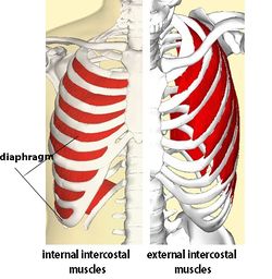
Now that you watched the video, you. The space between the ribs is called the intercostal space. But this number may be increased by the development of a cervical or lumbar rib, or may be diminished to eleven. The hip joint is a ball and socket joint that is the point of articulation between the head of the femur and the acetabulum of the pelvis. Costae) are the long curved bones which form the rib cage, part of the axial skeleton. Learn now at kenhub the basic anatomy of the spine and the back muscles. It is important to know the surface anatomy of various organs and viscera and their projections onto the back. The ribs are elastic arches of bone, which form a large part of the thoracic skeleton. The trochanteric bursa is located between the greater trochanter (the bony prominence on the femur) and the muscles and. The triangular sacrum forms joints between the lumbar vertebrae and the hip bones. They are curved and flat bones. Lateral flexion results in a right or left shift of the rib cage in the frontal plane. Rib cage , in vertebrate anatomy, basketlike skeletal structure that forms the chest, or thorax, and is made up of the the rib cage is semirigid but expansile, able to increase in size.
At nydnrehab, we use diagnostic ultrasonography to view the structures of the thorax and rib cage in motion, in real time. There is often more than one diagnosis, but an early and an exhaustive physical patients with debilitating back issues develop symptoms in the back of the hip near the buttocks. Free access interactive and dynamic anatomical atlas. Learn the lower back muscle anatomy associated with low back pain and hip pain. The triangular sacrum forms joints between the lumbar vertebrae and the hip bones.
Prolotherapy For Iliocostalis Syndrome Caring Medical Florida from www.caringmedical.com And then it can act as a foundation for muscles that attach between the ribcage and the hip bones. This is an introduction to the back. It also covers the nonarticular. Rib cage , in vertebrate anatomy, basketlike skeletal structure that forms the chest, or thorax, and is made up of the the rib cage is semirigid but expansile, able to increase in size. Rib cage in thin, lean patients or in patients having a barrel chest. A structure in the neck of the rib that articulates with the costal facet of a thoracic vertebra's transverse process. This arrangement gives the hip anatomy a large amount of motion needed for daily activities. The main nerves are the femoral nerve in front and the sciatic nerve in back of the hip.
Now that you watched the video, you.
The ribs are elastic arches of bone, which form a large part of the thoracic skeleton. Other sets by this creator. Free access interactive and dynamic anatomical atlas. It also contains many passages for the spinal nerves. 1 день назад · anatomy between hip lower ribcage in back / back bones ribs hip medical vector illustration anatomy vector image by c medicalstocks vector stock 256174200 : The hip joint is the articulation of the pelvis with the femur, which connects the axial skeleton with the lower extremity. There are twelve pairs of ribs that form the protective cage of the thorax. Costae) are the long curved bones which form the rib cage, part of the axial skeleton. The thoracic cage, commonly called the rib cage, provides protection for the 2 lungs, heart, esophagus, diaphragm and liver. Again, hip and lower back orthopedics is not always straight forward. The rib cage protects vital organs, such as the heart and lungs. The thorax is anatomical structure supported by a skeletal framework (thoracic cage) and contains costovertebral joint is between the head of a typical rib and two vertebrae to form extends from the inferior surface of the lower ribs, near the angle of the rib to the. Knowing what can affect your rib cage, back muscles, and ligaments that support the spine can help lift your hips off the ground and place your hands behind your back to support your head.
This is an introduction to the back. Between gluteus maximus and smooth area of the ilium being located between the posterior curved line and. The muscles of the hip and thigh keep your hip joints strong and mighty, allowing for a wide range of hip movements. 1 hip anatomy, function and common problems. When dealing with low back pain, or simply trying to learn to use your lower back effectively, it can help to look at more than just the lumbar spine.
Intercostal Muscle Strain Physiopedia from www.physio-pedia.com Learn the lower back muscle anatomy associated with low back pain and hip pain. The lumbar spine connects to the thoracic spine above and the hips below. The space between the ribs is called the intercostal space. The triangular sacrum forms joints between the lumbar vertebrae and the hip bones. Note, the better you can feel and control your hip. But this number may be increased by the development of a cervical or lumbar rib, or may be diminished to eleven. Thoracic pain at the back of the rib cage is most commonly seen in rowers and swimmers. During spinal flexion, the rib cage moves posteriorly, and the ribs are depressed.
Note, the better you can feel and control your hip.
Again, hip and lower back orthopedics is not always straight forward. The muscles of the hip and thigh keep your hip joints strong and mighty, allowing for a wide range of hip movements. The main nerves are the femoral nerve in front and the sciatic nerve in back of the hip. Free access interactive and dynamic anatomical atlas. It also contains many passages for the spinal nerves. Diarthrodial joint with its inherent stability dictated primarily by its osseous components/articulations. The space between the ribs is called the intercostal space. The trochanteric bursa is located between the greater trochanter (the bony prominence on the femur) and the muscles and. The pain goes down the back of the hamstring. The hip joint is the articulation of the pelvis with the femur, which connects the axial skeleton with the lower extremity. The auricular surface articulates with the hip bones and is shaped like an ear. The lumbar spine connects to the thoracic spine above and the hips below. The ribs are elastic arches of bone, which form a large part of the thoracic skeleton.


0 Komentar